42 images of human heart with labels
Printable Muscle Labeling Worksheet - Math Worksheets Grade 5 We hope your happy with this Printable Anatomy Labeling Worksheets Label Muscles Worksheet with Images idea. Showing top 8 worksheets in the category - Label The Muscles Of The Body. Vf 4330 Eye Diagram To Label Printable Schematic Wiring from static-cdnimageservicecloud. There are over 1000 muscles in your body. January 17 2021 kids. How the Heart Works - The Heart | NHLBI, NIH Blood also carries carbon dioxide to your lungs so you can breathe it out. Inside your heart, valves keep blood flowing in the right direction. Your heart's electrical system controls the rate and rhythm of your heartbeat. A healthy heart supplies your body with the right amount of blood at the rate needed to work well.
Heart - Wikipedia The human heart is situated in the mediastinum, at the level of thoracic vertebrae T5-T8.A double-membraned sac called the pericardium surrounds the heart and attaches to the mediastinum. The back surface of the heart lies near the vertebral column, and the front surface sits behind the sternum and rib cartilages. The upper part of the heart is the attachment point for several large blood ...

Images of human heart with labels
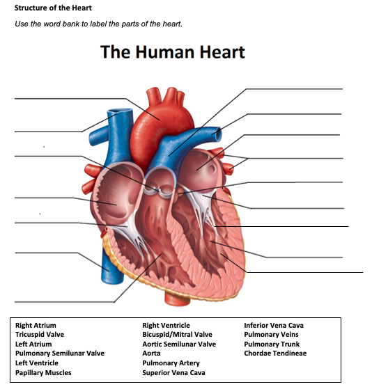
Anatomical Planes of Body - The Human Memory The X-axis is going from left to. right, Z-axis from front to back, and Y-axis from up to down. In anatomical. terminology, three references plane are considered standard planes; these. planes differentiate the body anterior and posterior, ventral and dorsal, dexter, and sinister portions. Let me tell you about these standard planes in detail. Diagram of Human Heart and Blood Circulation in It Exterior of the Human Heart A heart diagram labeled will provide plenty of information about the structure of your heart, including the wall of your heart. The wall of the heart has three different layers, such as the Myocardium, the Epicardium, and the Endocardium. Here's more about these three layers. Epicardium Reinforcement: Anatomy of the Human Heart Reinforcement: Anatomy of the Human Heart. I created this worksheet for my anatomy students to review the anatomy of the heart. Students focus on vocabulary that relates to anatomical features, such as the mitral valve, aorta, and heart chambers. At the end, students label a line drawing of the heart. I included a word bank at the top, but you ...
Images of human heart with labels. 40 Interesting Facts About Human Heart The heart is responsible for pumping blood to all 75 trillion cells present in human body. The only place where blood is not sent by the heart are the human corneas. 9. Heart is the only muscle in human body which does the maximum amount of physical work, generating around 1-5 watts of power. Even with 1 watt power, the heart can produce 2.5 ... Uyghur camps: Hacked info proof of China's secret detention system The data provides an unprecedented glimpse inside Beijing's internment of Uyghurs and other targeted ethnic groups. Zenz says he determined that at least 2,884 of the people in the photos were in ... Total Artificial Heart - What Is Total Artificial Heart? | NHLBI, NIH A total artificial heart (TAH) is a pump that is placed in the chest to replace damaged heart ventricles and valves. (Ventricles pump blood to the lungs and other parts of the body.) Once the pump has been placed in the chest, a machine called a driver controls the pump outside the body. The pump and driver help blood flow to and from the heart ... Heart Drawing With Label : Human Heart Diagram Images Stock Photos ... Heart Drawing With Label : Human Heart Diagram Images Stock Photos Vectors Shutterstock. Internal structure of human heart shows four chambers viz. Download this sketch of human heart anatomy with hand written labels vector illustration now. Two atria and two ventricles and couple of blood vessels opening into them.
Free Circulatory System Worksheets and Printables These worksheets will help your kids learn the parts of the heart as they label and color the different parts of the heart. Our Human Body Systems Labeling and Diagramming Worksheets have an Instant Download for the Circulatory System. This will help your children memorize the different parts of the circulatory system by using the free labels ... Layers of the heart: Epicardium, myocardium, endocardium - Kenhub The myocardium is functionally the main constituent of the heart and the thickest layer of all three heart layers. It is a muscle layer that enables heart contractions. Histologically, the myocardium is comprised of cardiomyocytes.Cardiomyocytes have a single nucleus in the center of the cell, which helps to distinguish them from skeletal muscle cells that have multiple nuclei dispersed in the ... What Are the Four Main Functions of the Heart? - MedicineNet The heart is a muscular organ situated in the chest just behind and slightly toward the left of the breastbone. It roughly measures the size of a closed fist. The heart works all the time, pumping blood through the network of blood vessels called the arteries and veins. The heart and its blood vessels are known as the cardiovascular system. Human Heart for Kids: 2 Fun Heart Models plus Worksheets Human Heart Coloring Pages - Identify the major parts of the human heart by coloring by code to identify the: Aorta, Left Atrium, Left Ventricle, Pulmonary Artery, Right Atrium, and Right Ventricle Human Body Activities for Kids Finally, we made a working heart pump model. As you can guess, this was the kids' favorite part.
Anatomy of the heart and coronary arteries (coronary CT) - IMAIOS Anatomy of the human heart and coronaries: how to view anatomical structures. This tool provides access to an MDCT atlas in the 4 usual planes, allowing the user to interactively discover the heart anatomy. The images are labeled, providing an important medical and anatomical tool. The quiz mode makes it possible to evaluate the user's progress. Diagrams, quizzes and worksheets of the heart - Kenhub Take a look at our labeled heart diagrams (see below) to get an overview of all of the parts of the heart. Once you're feeling confident, you can test yourself using the unlabeled diagrams of the parts of the heart below. Labeled heart diagram showing the heart from anterior Unlabeled heart diagrams (free download!) Automated multilabel diagnosis on electrocardiographic images ... - Nature The class mean weighted AUROC across clinical labels on the held-out test set images in both standard and alternate format was 0.99 (Supplementary Table 3). The class mean weighted AUPRC across ... Heart Diagram Grade 9 : Heart Cie Igcse Biology Revision Notes ... Grade 9 science students dissected pig hearts to understand the inner. Blood circulation, heart chambers, coronary arteries and other components of a healthy heart from froedtert. Unlike normal muscles the heart is made of cardiac muscle. Source: images.twinkl.co.uk. Heart diagram of coronary artery function .
HEART
71 Interesting Facts About Human Heart - The Fact File The superior vena cava and the inferior vena cava are the two largest veins that carry blood into the heart. 15. The heart receives blood which is low in oxygen and then the blood passes through the lungs where it is oxygenated. This oxygen-rich blood again enters the heart and is then sent to the body. 16.
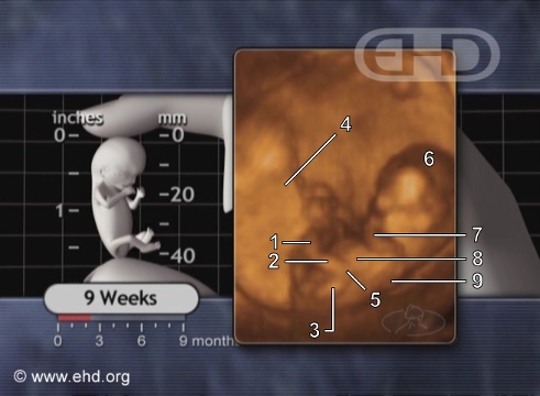
The 9-Week Fetus in Motion
Human heart: Anatomy, function & facts | Live Science An illustration of inside a human heart, showing all four chambers. (Image credit: Getty Images ) The heart's outer wall consists of three layers. The outermost wall layer, or epicardium, forms the...

The heart: How Does The Heart Work?
How the Heart Works: Diagram, Anatomy, Blood Flow - MedicineNet Illustrations of Blood Flow to the Heart Location and size of the heart The heart is located under the rib cage -- 2/3 of it is to the left of your breastbone (sternum) -- and between your lungs and above the diaphragm. The heart is about the size of a closed fist, weighs about 10.5 ounces, and is somewhat cone-shaped.

Simply Creative: Animal Body Art
Heart: illustrated anatomy - e-Anatomy - IMAIOS This interactive atlas of human heart anatomy is based on medical illustrations and cadaver photography. The user can show or hide the anatomical labels which provide a useful tool to create illustrations perfectly adapted for teaching. Anatomy of the heart: anatomical illustrations and structures, 3D model and photographs of dissection.

Human Anatomy Lab: The Urinary and Reproductive Systems
Anatomy of The Human Ribs - With Full Gallery Pictures! The Anatomy of the Human Ribs (costae) are one of the integral parts of the chest wall; they make up the lateral part of our body, its anterior and posterior wall and they entirely build the lateral parts of the chest wall. The anatomy of the human ribs is made up of 24 ribs. These ribs are parted in 12 pairs (each on the left and right side of ...

Lable Heart : New users enjoy 60% off. - Magic Pau
DailyMed - IBUPROFEN tablet temporarily relieves minor aches and pains due to: headache - muscular aches - minor pain of arthritis - toothache - backache - the common cold - menstrual cramps - temporarily reduces fever. Allergy alert: Ibuprofen may cause a severe allergic reaction, especially in people allergic to aspirin. Symptoms may include: hives - facial swelling ...

Human Anatomy Lab: Heart Models
Know Where Your Heart Is and How to Identify Heart Pain Here we are going to discuss the symptoms of several chest pains which are associated with heart. 1. Heart Attack. Heart attack results from the occluded blood vessels that carry blood to the heart. The patient may experience the following signs: Fullness or squeezing sensation in the chest.

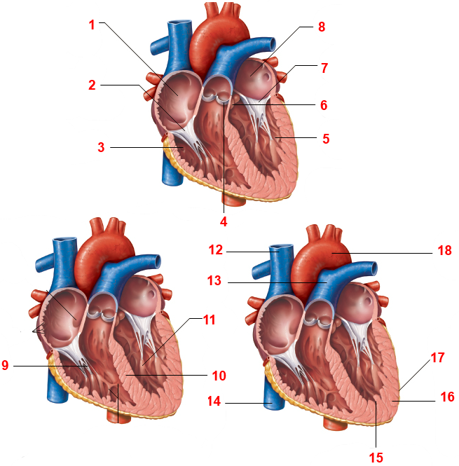
Identify Various Parts Of A Human Heart: Trivia Quiz - ProProfs Identify Various Parts Of A Human Heart: Trivia Quiz. The heart is the most important organ in the body. It is in charge of keeping the processes within the body moving by facilitating the transfer of blood throughout the body. The quiz below is to test out interesting facts you may know about the heart. Give it a try and good luck. 1. What is F?
31 Human Heart To Label - Labels Design Ideas 2020
Heart Labeling Quiz: How Much You Know About Heart Labeling? Here is a Heart labeling quiz for you. The human heart is a vital organ for every human. The more healthy your heart is, the longer the chances you have of surviving, so you better take care of it. Take the following quiz to know how much you know about your heart. Questions and Answers 1. What is #1? 2. What is #2? 3. What is #3? 4. What is #4?

Gaseous exchange in the lungs | Circulatory and respiratory systems | Siyavula
Free Heart Worksheets for Human Anatomy Lessons Print out sheet of the human heart with labels - This fun heart worksheet shows kids the different parts of the heart. They'll learn about the left ventricle, the left atrium, the tricuspid valve, and more. Human Heart Clipart - There is a coloring page, heart labeling worksheet and heart anatomy chart.

INFORMATION ABOUT HUMAN BODY.: THE HEART
Reinforcement: Anatomy of the Human Heart Reinforcement: Anatomy of the Human Heart. I created this worksheet for my anatomy students to review the anatomy of the heart. Students focus on vocabulary that relates to anatomical features, such as the mitral valve, aorta, and heart chambers. At the end, students label a line drawing of the heart. I included a word bank at the top, but you ...

Label the Heart Quiz
Diagram of Human Heart and Blood Circulation in It Exterior of the Human Heart A heart diagram labeled will provide plenty of information about the structure of your heart, including the wall of your heart. The wall of the heart has three different layers, such as the Myocardium, the Epicardium, and the Endocardium. Here's more about these three layers. Epicardium

Leadership: Group Work Can Be As Successful As You Want It To Be
Anatomical Planes of Body - The Human Memory The X-axis is going from left to. right, Z-axis from front to back, and Y-axis from up to down. In anatomical. terminology, three references plane are considered standard planes; these. planes differentiate the body anterior and posterior, ventral and dorsal, dexter, and sinister portions. Let me tell you about these standard planes in detail.
Heart - Human Body - Find Fun Facts
Labeling the Heart (Part Three) Quiz - By dilatory

The brain - structure and function - Cancer Information - Macmillan Cancer Support

Heart And Labels Drawing at GetDrawings.com | Free for personal use Heart And Labels Drawing of ...

Label the Heart - PurposeGames
Post a Comment for "42 images of human heart with labels"